
Figure 4. EphB2 forward signaling maintains the ependymal cell phenotype.
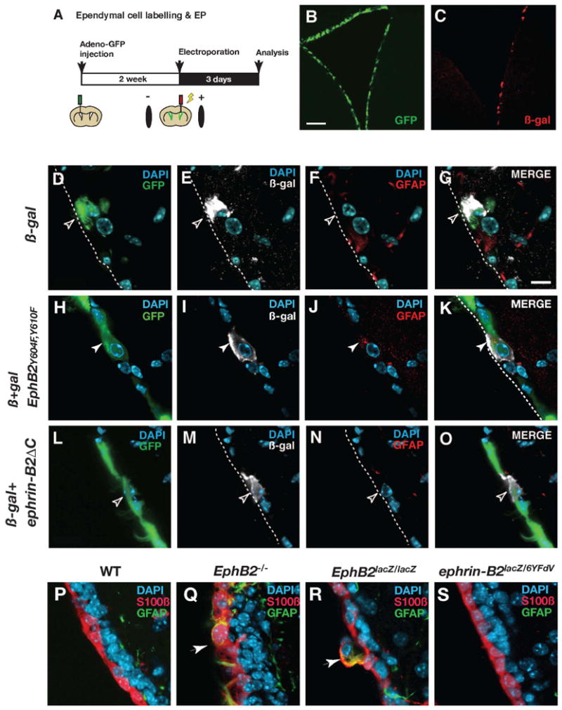
(A) Illustration of the experimental procedure. After labeling ependymal cells with adeno-GFP virus, plasmid vectors were electroporated into the lateral wall. (B and C) GFP expression by adenovirus transduction (B) and ß-galactosidase (ß-gal) expression by plasmid electroporation (C). (D-G) In a control animal electroporated with ß-gal expression vector, a cell which is double-positive for GFP and ß-gal does not express GFAP (open arrowheads). (H-K) In an animal electroporated with a vector expressing EphB2Y604F,Y610F together with the ß-gal expression vector, a cell which is double-positive for GFP and ß-gal expresses GFAP in the ependymal layer (white arrowheads). (L-O) In an animal electroporated with a vector expressing ephrin-B2ΔC together with the ß-gal expression vector, a cell which is double-positive for GFP and ß-gal does not express GFAP (open arrowheads). (P-S) Analysis of the ependymal layer in EphB2 or ephrin-B2-deficient mice. There are no GFAP and S100ß co-expressing cells in the ependymal layer of wild type mice (WT, P), but they are present (indicated by arrows) in both EphB2-/- (Q) and EphB2lacZ/lacZ mice (R). The ventricle wall appears indistinguishable from the WT in ephrin-B2lacZ/6YFdV mice (S). The luminal side of the ependymal layer is indicated by broken lines. Scale bars indicate 200 μm in (B) and 10 μm in (D).