
Figure 5.
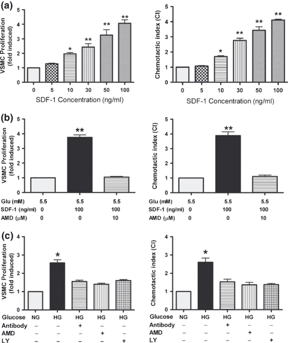
High glucose (HG) induced proliferation and chemotaxis of VSMCs. VSMCs were cultured in normal glucose (NG) medium containing 0, 5, 10, 30, 50 or 100 ng/ml of exogenous rat SDF-1α (a) or in NG medium supplemented with/without exogenous rat SDF-1α (100 ng/ml) and the CXCR4 antagonist, AMD3100 (AMD, 10 μM) (b) or in medium containing NG or HG supplemented with or without LY294002 (LY, 20 μM), AMD3100 (AMD, 10 μM), or neutralizing antibody against SDF-1α (Ab, 1 μg/ml) (c). Results are mean ± SEM of three independent experiments. *P < 0.05, **P < 0.01 (in A, vs. control only).