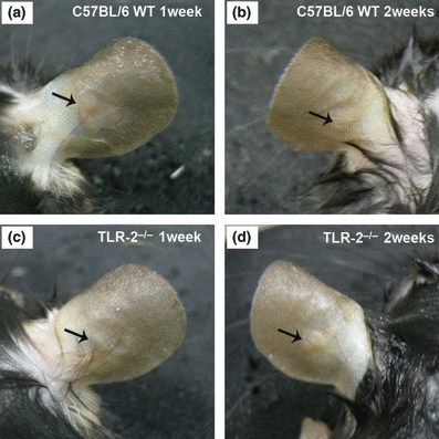
Figure 1.

Ear lesions in C57BL/6 WT and TLR-2−/− mice 1 and 2 weeks following intradermal inoculation of 2.5 × 10⁵ Leishmania (L.) amazonensis promastigotes. After the first week, C57BL/6 WT (a) and TLR-2−/− mice (c) present increased vascularization of the inoculation site. After the second week of infection, C57BL/6 WT mice (b) presented lesions with little ulceration, while in TLR-2−/− mice (d) the formation of small non-ulcerative nodular lesions were observed.
