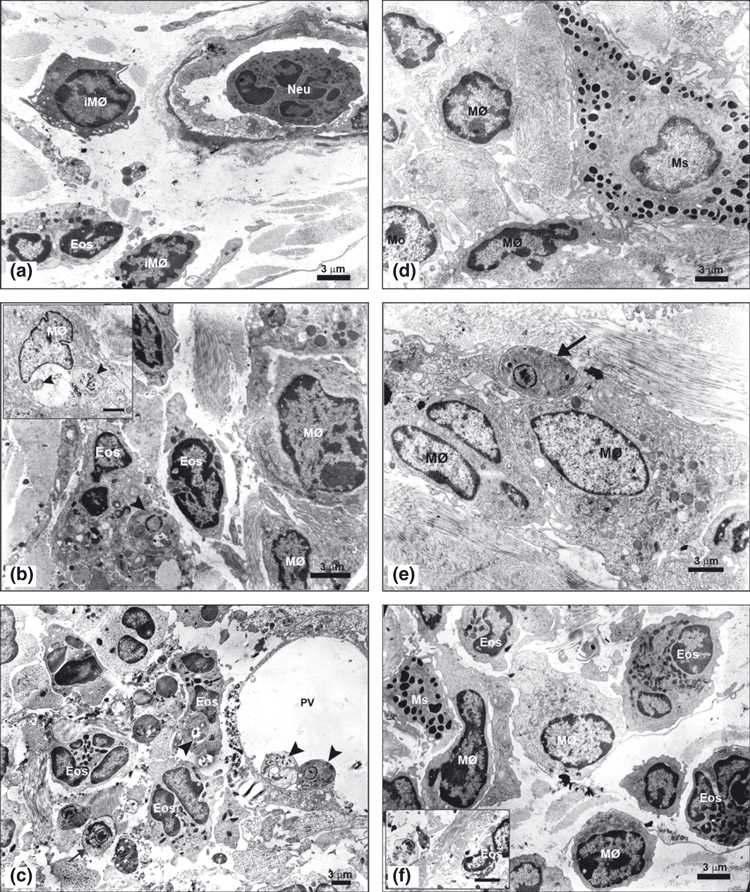
Figure 5.

(a–f) – Ultrastructural analysis of the ultra-thin sections from the ear lesion of the C57BL/6 WT and TLR-2−/− mice after 1, 7 and 15 days of Leishmania (L.) amazonensis infection contrasted with 5% uranyl acetate and lead citrate (bar = 3μm). Electron micrography of the first day infection (a) showed neutrophils (Neu) adhered to the endothelium of blood vessel, immature macrophages (iMØ) and eosinophils (Eos) in dermal ear of the C57BL/6 WT. After first week of the infection (b), eosinophils (Eos) containing one amastigotes within parasitophorous vacuoles close ( ) and immature macrophages characterized by few organelles and a nucleous with electron-dense chromatin were seen. Also, mature macrophages (MØ) containing amastigotes within large parasitophorous vacuoles were observed (inset). After the second week (c) mature macrophages (MØ) presented amastigotes (
 ) attached at the large parasitophorous vacuoles membrane (PV), many eosinophils (Eos) parasitized contained only one amastigote within parasitophorous vacuoles close and free amastigotes (
 ) in extracellular matrix were found. Furthermore, several biconvex granules, proceeding from eosinophils, were observed free in the extracellular matrix. In the first day of TLR-2−/− mice infection (d) were found mast cells (Ms) with electron-dense granules characteristic of this cell, distributed in the cytoplasm and macrophages (MØ). In the first week (e), presented some free amastigotes (
 ) in the extracellular matrix showed entire plasma membrane and nucleous with chromatin attached to the nuclear envelope and central nucleolus being phagocytized by mature macrophages (MØ). And, in the second week (f), mature macrophages (MØ), eosinophils (Eos) and mast cells (Ms) in ear dermis were observed. In this time, rare free amastigotes in the extracellular matrix presented cytoplasm rarefied and disruption at the plasma membrane and nuclear envelope, indicating cell death of these parasites (inset). The experiment is representative of three separate experiments.
