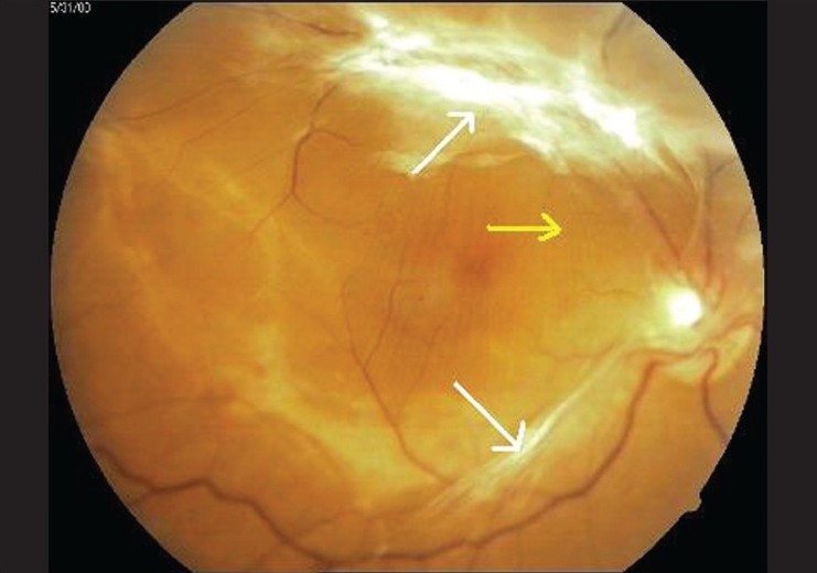
Figure 1.

Fundus photograph right eye - fibrovascular traction bands extending from optic disc, along both superior and inferior temporal arcades (white arrows) with taut posterior hyaloid membrane (yellow arrow) leading to folds in internal limiting membrane
