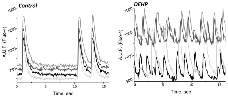
Figure 1. Physiological changes in cardiomyocyte behavior caused by DEHP (50 μg/mL) treatment.
(A) Calcium transient measurements were recorded from 4 regions of interest on a cardiomyocyte monolayer using Fluo-4, a calcium indicator dye. DEHP treatment (50 μg/mL) results in marked uncoupling between the different regions of the cell network (right) compared with untreated control samples (left).

