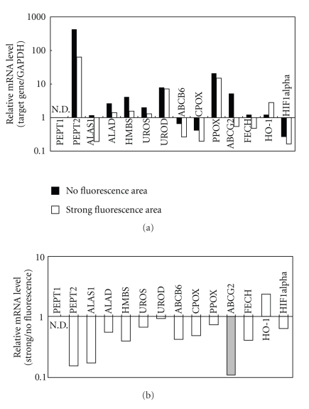
Figure 2.
The mRNA levels of PEPT1, PEPT2, ALAS1, ALAD, HMBS, UROS, UROD, ABCB6 CPOX, PPOX, ABCG2, FECH, HO-1, and HIF1alpha in malignant glioma cells in the brain tumor and in the surrounding normal cells. Total RNA was extracted from strong fluorescence-emitting areas (brain tumor) as well as from the surrounding area (mostly normal tissue) without porphyrin fluorescence. The first strand cDNA was prepared from the extracted total RNA in a reverse transcriptase (RT) reaction. (a) The mRNA levels of the genes involved in the heme synthesis and metabolism reactions were detected by quantitative PCR and normalized to the mRNA level of GAPDH. (b) The relative mRNA levels of those genes in both areas were compared, where the relative mRNA level in the strong fluorescence-emitting area (brain tumor) was divided by that in nonfluorescence area (mostly normal tissue).

