Figure 5.
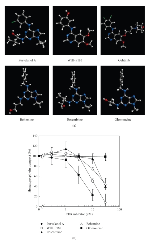
(a) Chemical structures of CDK inhibitors: purvalanol A, WHI-P180, gefitinib, bohemine, roscovitine, and olomoucine. (b) Inhibition of ABCG2-mediated hematoporphyrin transport by purvalanol A, WHI-P180, roscovitine, bohemine, or olomoucine. ABCG2-expressing plasma membrane vesicles (20 μg of protein) were incubated with 20 μM hematoporphyrin in the presence of purvalanol A, WHI-P180, roscovitine, bohemine, or olomoucine (final concentration: 0, 0.3, 1, 3, 10, or 30 μM) in the standard incubation medium (0.25 M sucrose and 10 mM Tris/HEPES, pH 7.4, 1 mM ATP, 10 mM creatine phosphate, 100 μg/mL of creatine kinase, 10 mM MgCl2) at 37°C for 10 min. Hematoporphyrin transported into membrane vesicles was detected [43]. Data are expressed as mean values ± SD (n = 5).

