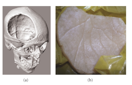
Figure 1.
(a) Three-dimensional reconstruction of skull defect based on computed tomography (CT) scans of a patient who had a right frontoparietal craniectomy. (b) The arborizing course of the anterior branch of the middle meningeal artery can be observed on the internal aspect of the bone flap, providing a model for design of vascular channels.

