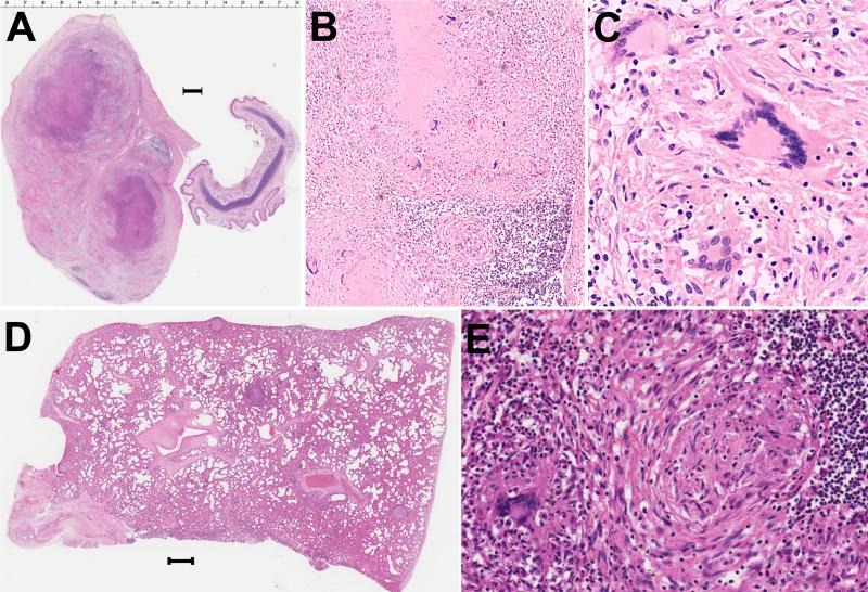
Figure 2.
A) One of the large tracheal-bronchial lymph nodes with caseation necrosis and the smaller trachea for comparison. Bar is 1 mm. 1 x, H&E. B) Histology of hilar lymph node showing amorphous necrosis. 100 x, H&E. C) Giant cells within the granulomas of the hilar lymph node, 400 x, H&E.. D) Lung with multiple granulomas throughout the parenchyma and at pleural surface. Bar is 1 mm. 1x, H&E. E. Histology of lung granuloma. 200x, H&E.

