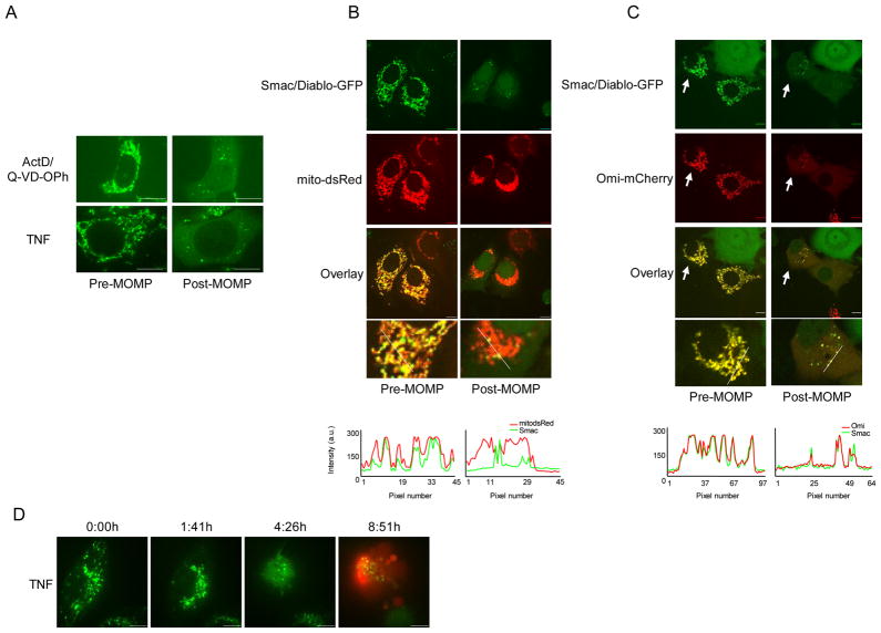
Figure 2.
Incomplete MOMP is cell-type and caspase independent. (A) MCF-7 cells expressing Smac-GFP were treated with UV (18mJ/cm2) plus Q-VD-OPh (20μM) or TNF (10ng/ml) and imaged every 10 minutes by live-cell confocal microscopy. Representative confocal micrographs pre- and post-MOMP are shown. (B) MCF-7 cells expressing Smac-GFP and matrix-targeted mCherry were treated with ActD (1μM) plus Q-VD-OPh (20μM) and imaged every 10 minutes by live-cell confocal microscopy. Representative confocal micrographs pre- and post-MOMP are shown. Line scans indicate co-localization of Smac-GFP and mito dsRed and correlate to the line drawn in the images. (C) MCF-7 cells expressing Smac-GFP and Omi-mCherry were treated with ActD (1μM) plus Q-VD-OPh (20μM) and imaged every 10 minutes by live-cell confocal microscopy. Representative confocal micrographs pre- and post-MOMP are shown and arrows denote cells undergoing iMOMP. (D) HeLa cells expressing Smac-GFP were treated with TNF (10ng/ml) in the presence of propidium iodide (1μg/ml) and imaged every 10 minutes by live-cell confocal microscopy. Representative confocal micrographs pre- and post-MOMP are shown. Scale bars represent 10μm. See also Figure S2.

