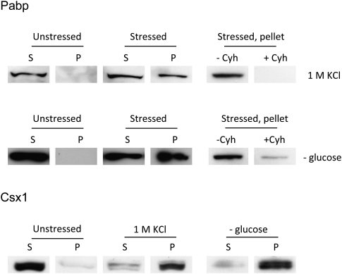
FIGURE 4.
Pabp and Csx1 are enriched in the insoluble protein fraction after stress. Wild-type cells expressing Pabp-RFP or Csx1-GFP were grown to mid-log phase and exposed to 1 M KCl for 30 min or deprived of glucose for 20 min. Protein lysates from equal numbers of stressed and unstressed cells were prepared and the protein concentration determined by reading absorbance at 280 nm. Equal protein amounts were then subjected to sequential centrifugations as described in Materials and Methods. Pellets (P) formed by centrifugation and samples from supernatant (S) were prepared for Western blot analysis; 10% of each supernatant, without further concentration, and 100% of each pellet fraction, after solubilization, was applied to the gel. Pabp was detected with anti-RFP (Genescript) and Csx1 with anti-GFP (Santa Cruz Biotechnology). Cycloheximide (Cyh) was added to 100 μg/mL before application of stress in some samples to demonstrate prevention of the insoluble fraction (right panels).

