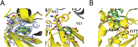
FIGURE 4.
Recognition of the branch nucleotide base during spliceosome assembly. (A) X-ray structure of adenine bound to p14/SF3b155 peptide. (Left) 2Fo − Fc map at 2.4 Å resolution using phases calculated from the final, refined model and contoured at 1σ (blue) and Fo − Fc map using phases calculated from a model lacking adenine contoured at +5σ (green). (Right) Interactions between adenine and the SF3b14 pocket. The purine stacks on Y22 of RNP2, features Watson-Crick-like hydrogen-bonding interactions between the N-6 exocylic amine and the main-chain carbonyl of Y91 and between N-1 and the main-chain N-H of N93, as well as hydrogen-bonding between N-3 and the hydroxyl of otherwise buried Y61 of RNP1. (B) Recognition of human branch region by the KH domain of SF1. Detail from the NMR structure showing specific recognition of the branch nucleotide base and SF1 mediated by Watson-Crick like interactions with the main chain of I177 (Liu et al. 2001).

