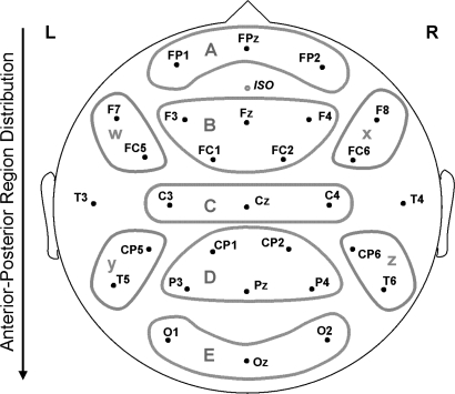
Fig. 1.
Electrode Montage and Regions Used in Analysis. Midregions: A, anterior frontal; B, frontal; C, central; D, central posterior; E, posterior. Peripheral regions: W, left anterior; X, right anterior; Y, left posterior; Z, right posterior. This and all other figures are available in color as online supplementary material.

