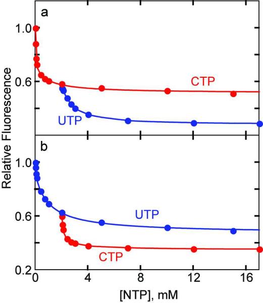
Figure 5.
Interaction of ATCase with CTP and UTP. a) Alteration in the fluorescence of the HCE-GLy52R ATCase at pH 8.3 upon the binding of UTP to the enzyme•CTP complex. The red circles represent saturation with CTP only, and the blue circles represent saturation with UTP in the presence of 2 mM CTP. b) Alteration in the fluorescence of the HCE-Gly52R ATCase upon the binding of CTP to the enzyme•UTP complex. The blue circles represent saturation with UTP only, and the red circles represent the saturation with CTP in the presence of 2 mM UTP. 50 μg mL−1 of HCE-Gly52R ATCase in 50mM Tris-acetate buffer at pH 8. at 25 °C was excited at 360 nm. The fluorescence intensity at 455 nm was normalized to the first data point (0 mM NTP) and plotted versus the nucleotide concentration.

