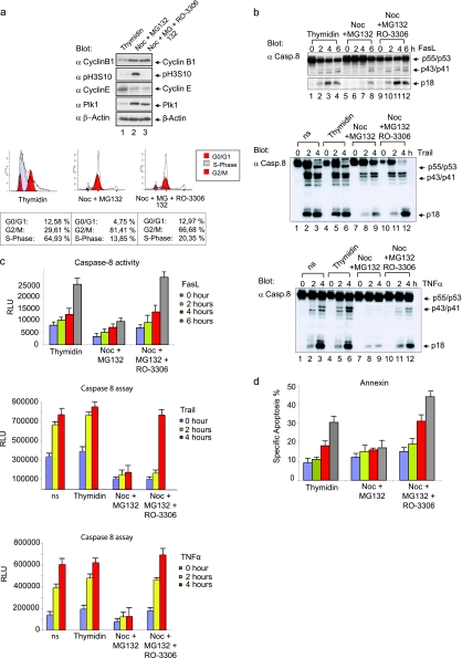
FIG. 1.
Inhibition of the extrinsic death pathway in Fas-induced mitotic cells. (a) B lymphoblastoid SKW6.4 cells were enriched in G1/S phase by treatment with thymidine (lane 1), in pro-/metaphase with nocodazole for 16 h, followed by MG132 for 2 h (lane 2) or in a G1-like state with nocodazole for 16 h, followed by MG132 for 2 h and RO-3306 for the inhibition of Cdk1/cyclin B1 (lane 3) (upper panel). Cell lysates were immunoblotted for cyclin B1, histone H3 phosphorylated at Ser10 (H3 pS10), cyclin E, Plk1, and β-actin. The cell cycle status was monitored by flow cytometry (lower panel). (b) Cells treated as described in panel a were stimulated with 100 ng of FasL/ml, 10 ng of TRIAL/ml, or 10 ng of TNF-α/ml for the times indicated, and total cellular lysates were analyzed by Western blotting with anti-caspase-8 MAb C15. (c) Caspase-8 activity was determined by measuring the cleavage of the luminogenic substrate containing the IETD peptide. All experiments were performed in triplicate. Error bars represent the standard deviation (SD). (d) Apoptosis analyses were performed by using an annexin V kit. All experiments were performed in triplicate. Error bars represent the SD.

