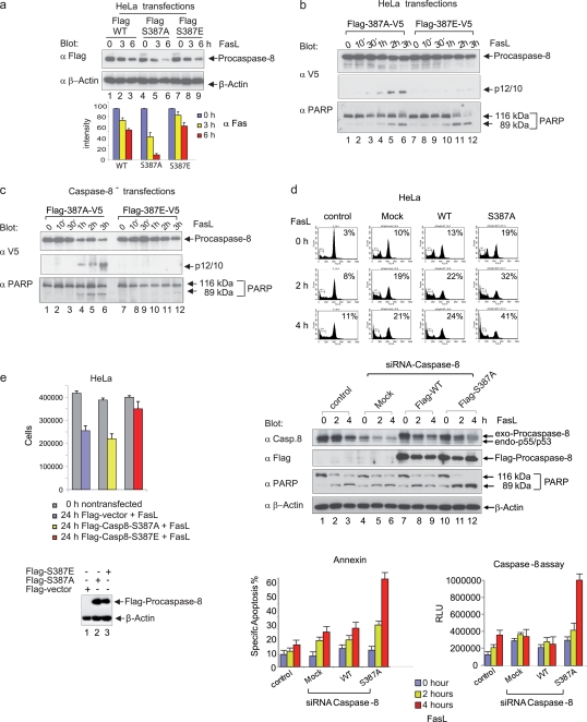
FIG. 7.
A nonphosphorylatable S387 mutant of procaspase-8 sensitizes mitotic cells to Fas-mediated apoptosis. (a) HeLa cells were transiently transfected with vectors encoding Flag-tagged caspase-8 (Flag-WT, Flag-S387A, and Flag-S387E). At 18 h after transfection, cells were incubated with 100 ng of FasL/ml plus 1 μg of cycloheximide/ml for 3 and 6 h. Lysates were immunoblotted for exogenous caspase-8 with a Flag-specific antibody and for β-actin (upper panels). The signal intensity of immunoblotted, Flag-tagged caspase-8 was standardized to the level of β-actin expression (lower panel). All experiments were performed in triplicate. The error bars represent the SD. (b) Analysis of procaspase-8 processing in HeLa cells expressing different double-tagged (N-terminal Flag, C-terminal V5) forms of procaspase-8 (S387A, S387E). Lysates were immunoblotted for V5 and PARP. (c) Analysis of procaspase-8 processing in caspase-8 knockdown HeLa cell clones (caspase-8−) expressing different double-tagged (N-terminal Flag, C-terminal V5) forms of procaspase-8 (S387A, S387E). Lysates were immunoblotted for V5 and caspase-8. (d) On day 1, HeLa cells were transfected with siRNA targeting the untranslated region of caspase-8, followed by the transfection of Flag-tagged caspase-8 (mock, WT, or S387A) on day 2. On day 3, the cells were treated overnight with nocodazole, and then a mitotic shake-off was performed on day 4. Subsequently, cells were reseeded in nocodazole-containing medium and stimulated with 100 ng of FasL/ml. The cell cycle status was monitored by flow cytometry (upper panels). Lysates were immunoblotted for exogenous caspase-8 using a Flag-specific antibody, caspase-8, PARP, and β-actin (middle panels). Caspase-8 activity and apoptosis (annexin staining) were determined (lower panels). All experiments were performed in triplicate. Error bars represent the SD. (e) On day 1 HeLa cells were transfected with siRNA targeting the untranslated region of caspase-8, followed by the transfection of Flag-tagged caspase-8 (mock, S387A, or S387E) on day 2. On day 3 cells were stimulated with 100 ng of FasL/ml. After 24 h, the cell numbers were determined, and lysates were immunoblotted for exogenous caspase-8 using a Flag-specific antibody and β-actin. The experiment was performed in triplicate. Error bars represent the SD.

