FIG. 2.
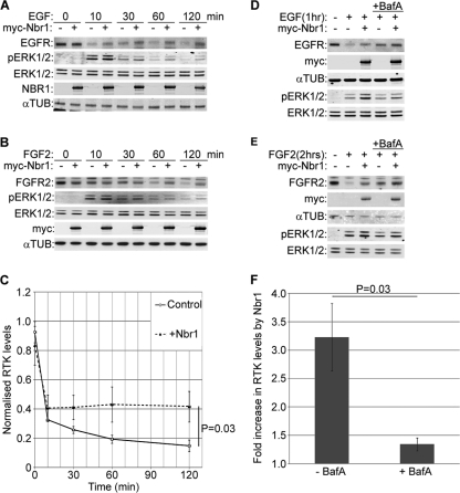
Nbr1 abrogates ligand-mediated lysosomal degradation of RTKs and enhances downstream signaling. All error bars represent standard errors of the means (SEM). P values were calculated by a one-tailed paired t test (n = 3). (A) Overexpression of Nbr1 inhibits ligand-mediated EGFR degradation and enhances downstream ERK1/2 signaling. HEK293T cells transfected with Myc-Nbr1 or GFP as a control were serum starved and stimulated for the indicated time points with 50 ng/ml EGF. Cells were lysed and analyzed by Western blotting. (B) Overexpression of Nbr1 inhibits ligand-mediated FGFR2 degradation and enhances downstream ERK1/2 signaling. HEK293T cells transfected with Myc-Nbr1 or GFP as a control were serum starved and stimulated for the indicated time points with 20 ng/ml FGF2 plus 10 μg/ml heparin. Cells were lysed and analyzed by Western blotting. (C) Densitometry analysis of RTK degradation in the presence or absence of Myc-Nbr1 from panels A and B. (D) Nbr1 inhibition of ligand-mediated EGFR degradation is BafA sensitive. HEK293T cells transfected with Myc-Nbr1 or GFP as a control were starved and stimulated for the indicated times with 50 ng/ml EGF. Thirty minutes prior to stimulation, the specified cells were pretreated with 200 nM BafA. Cells were lysed and analyzed by Western blotting. (E) Nbr1 inhibition of ligand-mediated FGFR2 degradation is BafA sensitive. HEK293T cells transfected with Myc-Nbr1 or GFP as a control were starved and stimulated for the indicated times with 20 ng/ml FGF2 plus 10 μg/ml heparin. Thirty minutes prior to stimulation, the specified cells were pretreated with 200 nM BafA. Cells were lysed and analyzed by Western blotting. (F) Densitometry analysis of the effect of BafA treatment on Myc-Nbr1-mediated inhibition of RTK degradation from panels D and E. αTUB, α-tubulin.

