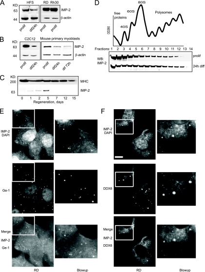
FIG. 1.
Expression levels and subcellular localization of IMP-2. (A to C) A Western blot shows that IMP-2 is expressed in human primary myoblasts (HFS) and in human RMS lines RD (embryonic) and Rh30 (alveolar) (A), in the mouse C2C12 cell line and primary myoblasts (B), and at the onset of skeletal muscle regeneration of mouse tibialis anterior muscle (C). (D) Sucrose gradient analysis of IMP-2 distribution between polysomal and monosomal fractions in C2C12 myoblasts. OD260, optical density at 260 nm. (E and F) IMP-2 forms distinct cytoplasmic granules and colocalizes with P-body markers Ge-1 and DDX6 in the RMS cell line RD. diff, differentiation; prolif, proliferation; DAPI, 4′,6-diamidino-2-phenylindole. Masses in kilodaltons are shown to the left of panels A to C. Bar, 9 μm.

