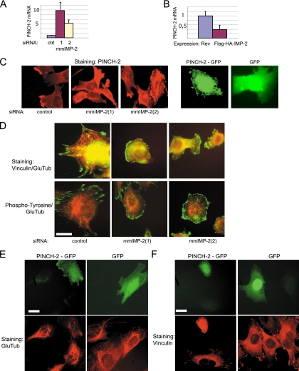
FIG. 3.
In C2C12 myoblasts, increased levels of PINCH-2 mRNA and protein lead to changes in cell shape, to clustering of FA structures, and to decrease in stable MTs. (A) KD of IMP-2 by two distinct siRNAs increases the levels of PINCH-2 mRNA. (B) Stable ectopic expression of IMP-2 decreases the levels of PINCH-2 mRNA. (C) Expression of PINCH-2 protein in IMP-2 KD C2C12 cells (left and middle panels) and in PINCH-2-GFP-transfected C2C12 cells (right panels); (D) immunofluorescent staining of FAs (green) and stable MTs (red) in control and IMP-2 KD cells; (E), decrease of Glu-tubulin staining in PINCH-2-GFP-expressing cells; (F) immunofluorescent staining of FAs in control and PINCH-2-GFP-expressing C2C12 myoblasts. Bars in panels D to F, 12 μm.

