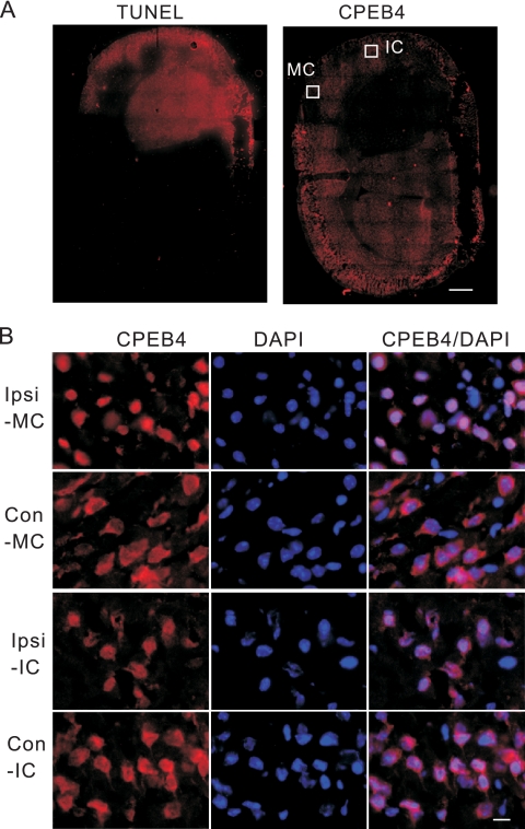
FIG. 6.
Ischemia causes CPEB4 protein to become concentrated in the nucleus. (A) A frozen section of brain taken from a mouse that had a middle cerebral artery occlusion (MCAO) performed was fixed and stained for CPEB4. A consecutive section from the same animal was labeled by TUNEL staining. The white boxes refer to regions of the motor cortex (MC) and insular cortex (IC) that were examined under higher magnification, as shown in panel B. Size bar = 1 mm. (B) A section from the ischemic brain was immunostained with anti-CPEB4 antibody. DAPI staining shows nuclei. The images were taken from the motor cortex (MC) or insular cortex (IC); ipsilateral (Ipsi) and contralateral sides (Con) of these regions are shown. Size bar = 20 μm.

