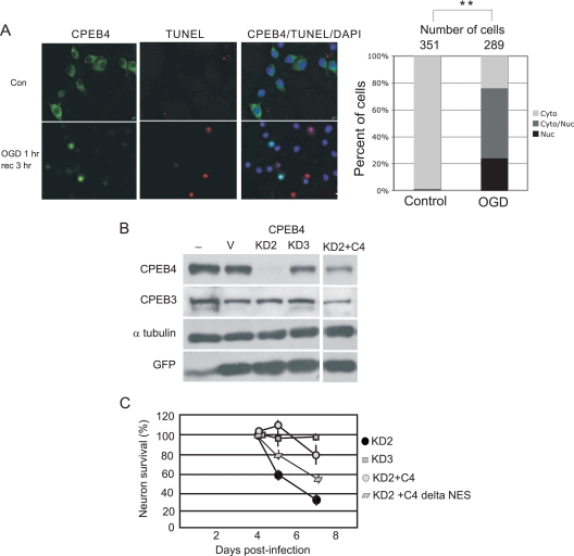
FIG. 7.
Oxygen and glucose deprivation and CPEB4-mediated neuron survival. (A) CPEB4 nuclear localization in DIV14 hippocampal neurons after OGD treatment. Hippocampal neurons were incubated in medium without glucose in an atmosphere deprived of oxygen for 1 h (OGD 1 h), which was followed by recovery in normal culture medium in atmosphere containing oxygen for 3 h (rec 3 h). Control (Con) refers to cells without OGD treatment. The images show CPEB4 staining, TUNEL staining, and CPEB4/TUNEL/DAPI staining to show the location of nuclei. Right, quantification of the CPEB4 proteins in the nucleus and cytoplasm as described in the legend to Fig. 1. The asterisks refer to a statistically significant difference (P < 0.01, Student's t test) between the results for the indicated samples. (B) Hippocampal neurons were cultured with lentivirus expressing GFP only (V) or GFP and two different CPEB4 shRNAs (KD2 and KD3). Some neurons were also cultured with two lentiviruses expressing KD2 and CPEB4 (C4), containing mutations to prevent knockdown but still encoding the correct protein. Extracts from the cells were probed for CPEB4, CPEB3, tubulin, and GFP. (C) Survival of the neurons infected with some of the viruses noted above was determined (n = 200). Error bars represent standard errors of the means.

