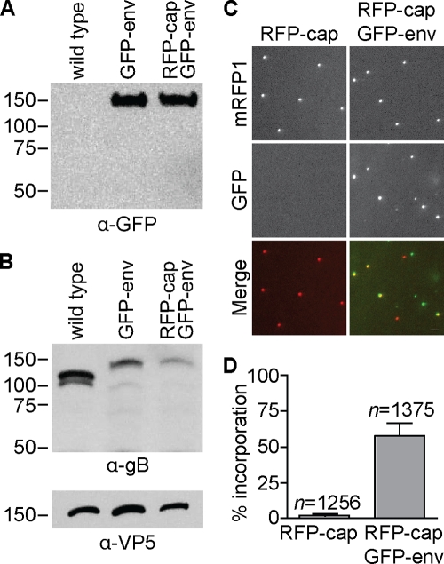
FIG. 3.
Incorporation of fluorescent fusion protein into extracellular viral particles. (A) Western blot of sucrose gradient-purified extracellular viral particles from the indicated HSV-1 strain probed with an anti-GFP antibody. (B) Western blot of sucrose gradient-purified extracellular viral particles from the indicated HSV-1 strains probed with anti-gB and anti-VP5 antibodies. The latter was included as a loading control. (C) Examples of GFP and RFP emissions from individual extracellular viral particles released from Vero cells 2 to 3 days postinfection with the indicated HSV-1 strain. The fields are each 24 μm by 24 μm; the bar is 2 μm. (D) GFP-gB incorporation frequency (GFP + RFP particles/total RFP particles) determined from images as represented in panel C. Averages from five individual experiments are shown, with error bars representing the standard deviation (n, number of capsid-containing particles imaged).

