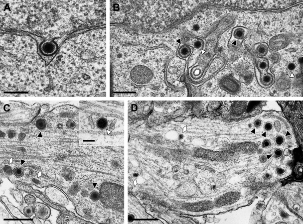
FIG. 1.
Ultrastructural analysis of primary rat neurons infected by HSV-1 HFEM. Micrographs show primary envelopment at the inner nuclear membrane (A), secondary envelopment in the cytosol (B), enveloped virions and neurovesicles (C) as well as a naked nucleocapsid (C, inset) in the axon, or enveloped virions and neurovesicles in the growth cone (D). Black triangles indicate enveloped virions; white triangles denote naked nucleocapsids. Neurovesicles are marked by lozenges. Bars: 200 nm in panel A, 300 nm in panel B, 500 nm in panels C and D, and 100 nm in panel C, inset.

