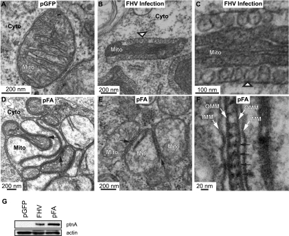
FIG. 2.
Expression of protein A causes mitochondrial zippering. (A) Electron micrograph of a cell expressing GFP showing no apparent alterations in mitochondrial morphology. (B) Electron micrograph of a cell infected with WT FHV at 14 hpi showing formation of mitochondrial spherules (arrowhead) that represent the FHV RNA replication complex. (C) Higher-magnification view of spherules in panel B. (D and E) Electron micrograph of cells expressing pFA, showing mitochondrial aggregation and zippering (arrows) but no spherules. (F) Higher-magnification view of zippered mitochondria, showing structure between mitochondria that may be responsible for zippering effect (arrows). Mitochondria (Mito), cytoplasm (Cyto), inner mitochondrial membrane (IMM), and outer mitochondrial membrane (OMM) are indicated. (G) Total protein was analyzed by Western blotting with antibodies against protein A (ptnA; top) or actin (bottom).

