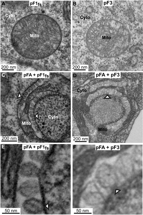
FIG. 6.
Coexpressing FHV RNA templates with WT protein A induces mitochondrial spherules. (A) Electron micrograph of a cell expressing pF1fs alone, showing no mitochondrial spherules. (B) Electron micrograph of a cell expressing pF3 alone, showing no mitochondrial spherules. (C) Electron micrograph of a cell expressing pFA plus pF1fs in which trans replication of RNA1fs is occurring (Fig. 5, lane 5), showing mitochondrial spherule formation (arrowheads). (D) Electron micrograph of a cell expressing pFA plus pF3 in which trans replication of RNA3 is occurring (Fig. 5, lane 7), showing mitochondrial spherule formation (arrowheads). (E and F) Higher-magnification views of spherules from panels C and D, respectively. Mitochondria (Mito) and cytoplasm (Cyto) are indicated.

