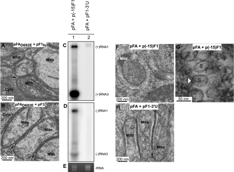
FIG. 7.
Spherule formation requires protein A polymerase activity and a replicatable RNA template. (A) Electron micrograph of a cell expressing pFAD692E plus pF1fs in which no replication is occurring (Fig. 5, lane 6), showing no mitochondrial spherule formation but mitochondrial zippering (arrow), as occurs when expressing pFA alone (Fig. 2D to F). (B) Electron micrograph of a cell expressing pFAD692E plus pF3 in which no replication is occurring (Fig. 5, lane 8), showing no mitochondrial spherule formation but mitochondrial zippering (arrows), as occurs in panel A and when expressing pFA alone (Fig. 2D to F). Drosophila cells were transfected with pFA plus an RNA1fs template with a truncation of the first 15 nt [p(−15)F1] or a deletion of the 3′ UTR [pF1-3′U]. Total RNA was analyzed by Northern blotting with 32P-labeled cRNA probes for positive-strand RNA1 and RNA3 (C) or negative-strand RNA1 and RNA3 (D). (E) EtBr-stained rRNA. (F) Electron micrograph of cells transfected with pFA plus p(−15)F1. (G) Higher-magnification view of spherules from panel F. (H) Electron micrograph of cells transfected with pFA plus pF1-3′U, showing mitochondrial zippering but no spherules. Mitochondria (Mito), cytoplasm (Cyto), and spherules (S) are indicated.

