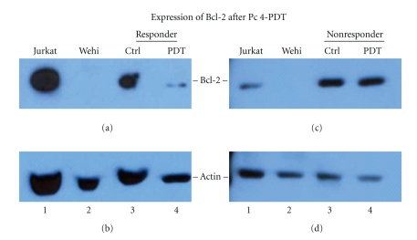
Figure 5.
Representative Western blots of Bcl-2 expression after in vivo treatment with Pc 4-PDT. Skin biopsies of MF lesions were obtained from patients diagnosed with MF. This Western blot compares Bcl-2 expression in a single representative clinical responder to that of a single representative clinical nonresponder. The top panels in both (a) and (c) show the 26 KDal Bcl-2. (b) and (d) show bands corresponding to actin as loading control. For both the responder (a and b) and nonresponder blots (c and d), Lane 1 consists of Jurkat cells, a cell line known to have endogenous expression of Bcl-2. Lane 2 consists of Wehi cells, a mouse cell line known to have minimal endogenous Bcl-2 expression. Lanes 3 and 4 contain the extraction from the untreated and treated lesions, respectively. (a) The clinical responder patient shows significantly decreased Bcl-2 expression in treated lesions in comparison to Bcl-2 expression in untreated lesion. In Lane 2, there is no corresponding band, as expected. (b) In the clinical nonresponder patient, again, there is Bcl-2 expression detected in Jurkat cells and no expression in Wehi cells, as expected. However, in contrast to the clinical responder patient, the level of Bcl-2 expression in treated and untreated lesions of the nonresponder is similar. These results are supported by the level of actin protein, which shows approximately similar amounts of protein loaded.

