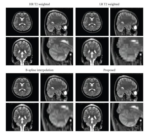
Figure 6.
Comparison on real clinical data. Top-Left: HR T2-w volume. Top-Right: downsampled version of the HR T2-w volume. Bottom-Left: B-spline reconstruction. Bottom-right: reconstruction using the proposed method. Note that the proposed methodology yields a significantly less blurred reconstruction than other methods compared. A close-up of the cerebellum area clearly shows the improved reconstruction.

