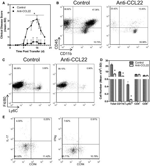
Figure 6. Anti-CCL22 treatment inhibits adoptively transferred EAE by reducing CNS inflammatory macrophages.
(A) Two groups of recipient mice (n=10) received i.v. injections of 5 × 10⁶ PLP139–151-primed and reactivated lymphocyte blasts and were treated with control or polyclonal anti-murine CCL22 at Days 4 and 6 relative to T cell transfer. The data represent the mean clinical disease score at each specific day post-cell transfer and significantly demonstrate (*P<0.05) disease development in the anti-CCL22-treated group. (B) The CNS of control- or anti-CCL22-treated mice was evaluated by flow cytometry for leukocyte infiltration. The results show a decreased percentage of lymphocytes (CD45hiCD11b–) and macrophages (CD45hiCD11b+) in a representative anti-CCL22-treated animal compared with control. (C) The CNS of control- or anti-CCL22-treated mice was evaluated by flow cytometry for inflammatory macrophage infiltration. The results show a decreased percentage of inflammatory macrophages (CD45hiF4/80+Ly6Chi) in a representative anti-CCL22-treated animal compared with control. (D) The number of CNS-infiltrating leukocytes was determined in control- and anti-CCL22-treated mice (n=5, each group). The results show a significantly (*P<0.05) decreased number of total leukocytes, macrophages, and Ly6Chi inflammatory macrophages in the CNS of anti-CCL22-treated mice. (E) CNS-infiltrating Th1 and Th17 cells from mice with EAE do not express CCR4. The results shown are representative of two similar experimental replicates.

