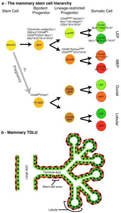
Fig. 1.
Developmental and functional hierarchies within the mammary gland. As described in the text, there is evidence of at least two potential stem cell hierarchies in the mammary gland. a Shows diagrams of the two putative mammary stem cell hierarchies. In one hierarchy, the quiescent mammary stem cells (MamSC) are thought to give rise to their nearest descendants, the bipotent progenitors (BPP), which subsequently give rise to luminal and myoepithelial line-age-restricted progenitors (LumRP and MyoRP, respectively). The lineage restricted progenitors subsequently generate differentiated ductal and luminal cells of the luminal epithelial (LEP) or myoepithelial (MEP) lineages. The second hierarchy originates from cells designated “parity-induced mammary epithelial cells” (PIMECs), which give rise to bipotent progenitors that generate both LEP and MEP, but are either duct- (ductal BPP) or lobule-specific (lobular BPP). Markers that have been used to describe or to isolate the different cell types are listed. Notably, CD24 and CD29 have so far only been used to identify murine mammary stem cells and have not yet been tested in human. b A cartoon of a terminal ductal-lobular unit is shown with color coding that corresponds to the stem cell hierarchy diagram. While the stem cell zone was reported in terminal ducts, the location of the BPP and lineage-restricted progenitors relative to the stem cells has yet to be described

