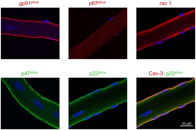
Figure 5. NADPH oxidase immunostaining of single fibers from mdx mice.
Isolated mdx muscle fibers from the FDB muscle were immunostained using antibodies against the various NADPH oxidase subunits. Nuclei are stained by DAPI (blue). In the bottom right panel, p22phox (green) was co-immunostained with caveolin-3 (red) to demonstrate sarcolemmal localization (yellow).

