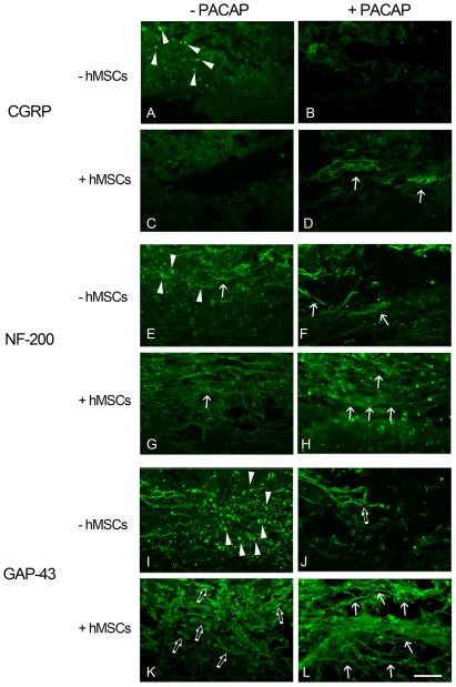
Figure 4. Immunofluorescence staining for identifying the remaining neuronal fibers in the lesion center of the injured spinal cord.
After BBB open field scores were performed at day 31, spinal cord tissues were collected. Longitudinal tissue sections were subjected to immunofluorescence for CGRP (A–D), NF-200 (E–H), or GAP-43 (I–L). CGRP positive neuronal fibers were observed in the dorsal section of the spinal cord (D, arrows). Elongated neuronal bundles labeled with CGRP, NF-200, or GAP-43 (arrows in D, H, and L) were observed in the injured spinal cord tissue receiving delayed combinatorial treatment of PACAP and hMSCs. Few NF positive or GAP-43 positive neuronal fibers were seen in the injured spinal cord tissues treated with PACAP (arrows in F and open arrows in G). Fine NF positive fibers (arrow in G) or fragmented GAP-43 positive fibers (open arrows in K) can be detected in the lesion center of the spinal cord with delayed treatment by hMSCs. Arrowheads indicate the cell debris with immunostaining for CGRP (A), NF-200 (E), or GAP-43 (I). Scale bar, 100 µm.

