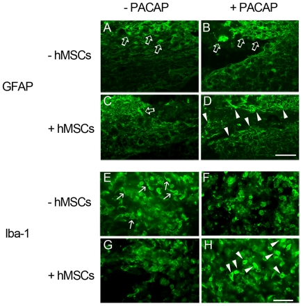
Figure 5. Immunofluorescence of GFAP and Iba-I in the injured spinal cord tissues. The injured spinal cord tissue sections were collected at day 31 after SCI.
The longitudinal tissue sections were subjected to immunofluorescence for GFAP (A–D) and Iba-1 (E–H). GFAP positive hypertrophic cells (open arrows) were observed in the areas proximal to the lesion center of the injured spinal cord treated with vehicle (A), PACAP (B), or hMSCs (C), while GFAP positive stellated cells (arrowheads) were found at the periphery to the lesion center treated with hMSCs and PACAP (D). Amoeboid-shaped Iba-1 positive microglia/macrophages (arrowheads) were observed in the injured spinal cord proximal to the lesion center with combinatorial treatment by hMSCs and PACAP (H). Unipolar Iba-1 positive microglia (arrows in E) are present around the lesion center of the spinal cord treated with vehicle. Scale bars, 100 µm (A–D) and 50 µm (E–H).

