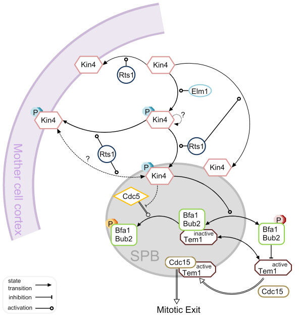
Figure 5.
Current model of SPOC activation. A simple molecular network of our current understanding of SPOC function. Details of the model can be found in the text. To which structural SPB component the proteins bind is not depicted in the figure and will be explained here: Bfa1-Bub2 associates with the SPBs through Nud1 [97]. Tem1 binding to the SPBs is via Bfa1-Bub2 except for during late anaphase [10]. Kin4 binds to Spc72 [84]. Cdc5 SPB localization largely depends on Cnm67 and Nud1 although it also interacts with Spc72 [95,108]. How Cdc15 localizes to the SPBs is not clear; however it depends on Tem1 and Cnm67 and to some extend Nud1 [91,96,97]. For simplicity only one SPB (outer plaque) has been depicted and all reactions are shown in one direction except for the highly dynamic SPB binding of Tem1 and Bfa1-Bub2 which is indicated by the two-sided arrows. However, Bfa1-Bub2 independent SPB binding of Tem1 in late anaphase was not depicted as a two-sided arrow for being a relatively more stable binding than the Bfa1-Bub2 dependent pool. Dashed-lines represent the potential reactions which are not known at the moment. Double-lines refer to the reactions that favor MEN activation. Hypothetical autophosphorylation of Kin4 is indicated by a question mark.

