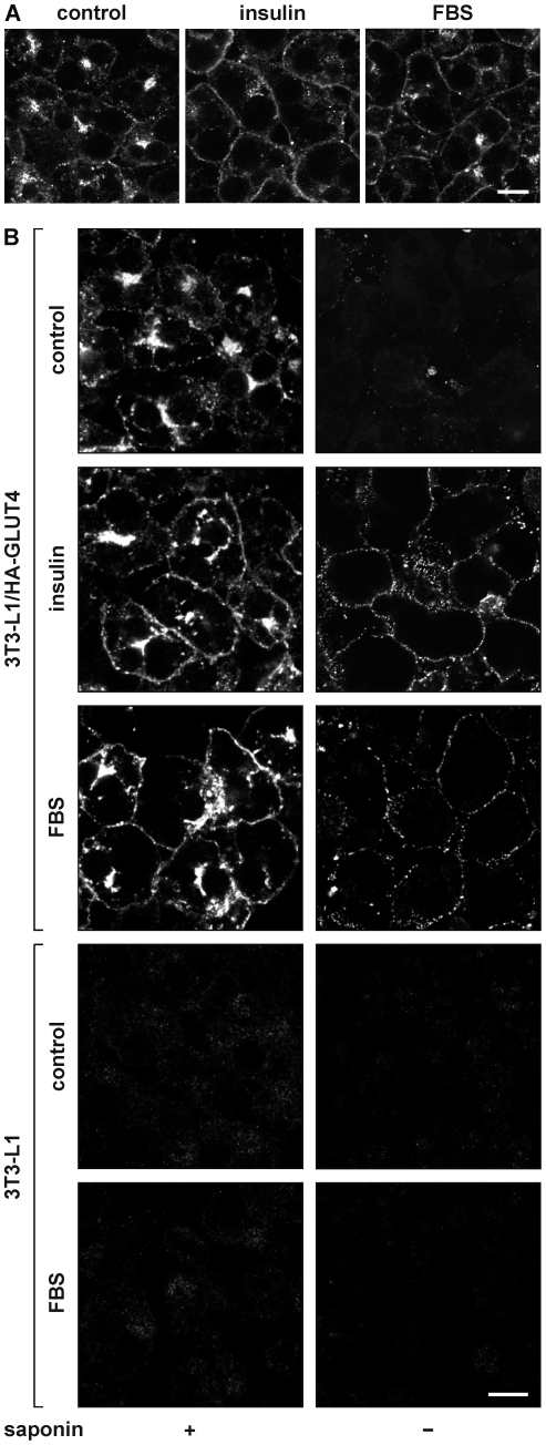
Figure 1. Effect of serum on the intracellular localization of GLUT4.
3T3-L1 adipocytes (A) or HA-GLUT4-expressing adipocytes (B) were incubated for 20 minutes with 100 nM insulin, 50% FBS, or left untreated (control). Upon fixation, cells were immunolabeled with anti-GLUT4 antibody to label endogenous GLUT4 (A) or with anti-HA in the absence (right panels) or presence of saponin (left panels) to label HA-GLUT4 at the cell surface or total cellular HA-GLUT4, respectively (B). Control adipocytes that did not express HA-GLUT4 were used to analyse the specificity of the anti-HA labeling (4 lower panels in B). Bar, 10 µm.

