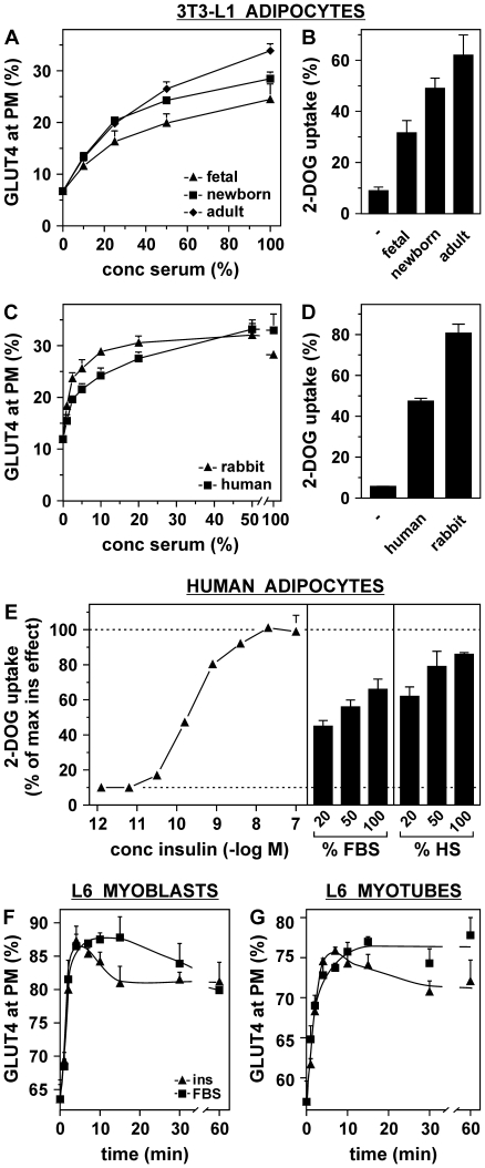
Figure 3. The effect of serum on GLUT4 is independent of serum origin and cell type.
(A) 3T3-L1 adipocytes were incubated for 20 minutes with the indicated concentrations of fetal, newborn, and adult bovine serum and cell surface GLUT4 levels were determined. (B) 3T3-L1 adipocytes were incubated for 20 minutes with 100 nM insulin, 50% serum or left untreated and cellular glucose uptake was measured and expressed as percentage of insulin-stimulated glucose uptake. (C, D) Rabbit and human sera were analyzed as under (A) and (B). (E) Human adipocytes were incubated for 20 minutes with various concentrations of insulin, FBS, or human serum (HS) and glucose uptake was measured and expressed as percentage of maximal glucose uptake in response to insulin. (F, G) HA-GLUT4-expressing myoblasts (F) and myotubes (G) were stimulated with either 100 nM insulin or 25% FBS and cell surface GLUT4 levels were determined. Serum insulin concentrations are depicted in Table S1.

