
Figure 4.
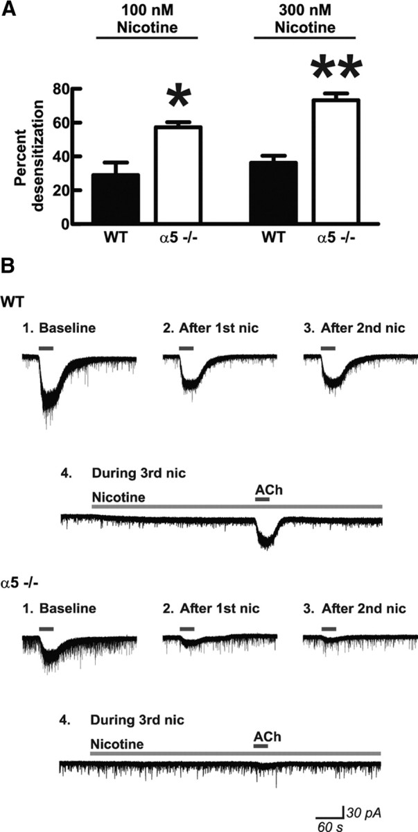
Presence of the nicotinic receptor α5 subunit protects medial prefrontal layer VI pyramidal neurons from nicotine-induced receptor desensitization. A, Nicotinic receptor desensitization by nicotine within individual neurons was calculated by measuring peak inward current responses to bath application of 1 mm acetylcholine (30 s) before and after exposure to either 100 or 300 nm nicotine for 10 min. There were significant effects of nicotine concentration (two-way ANOVA, p = 0.02) and genotype (p < 0.0001) on percentage desensitization after nicotine exposure. Neurons from mice in which the α5 subunit had been genetically deleted (α5−/−) showed greater desensitization compared with neurons from WT mice expressing the α5 subunit after exposure to both 100 nm nicotine (Bonferroni's post hoc test, *p < 0.01) and 300 nm nicotine (**p < 0.001). Values are mean ± 1 SEM. B, Example voltage-clamp traces in response to acetylcholine are shown before (Baseline) and after (After 1st nic) an initial 10 min exposure to 300 nm nicotine for one neuron each from a mouse of each genotype. Nicotine was washed out of the slices for 10 min, during which time current signals returned to baseline. Although there was an initial desensitization in neurons from WT mice, the resulting acetylcholine current response was preserved after a second 10 min exposure to nicotine (After 2nd nic) and was also preserved when acetylcholine was applied during a third 10 min exposure to nicotine (During 3rd nic). In contrast, the acetylcholine response was diminished or even abolished in neurons from α5−/− mice after subsequent 10 min exposures to nicotine. Note that nicotine continues to show activational effects (increase in inward current and current noise) even on the third application in the WT neuron but does not show these effects in the α5−/− neuron.