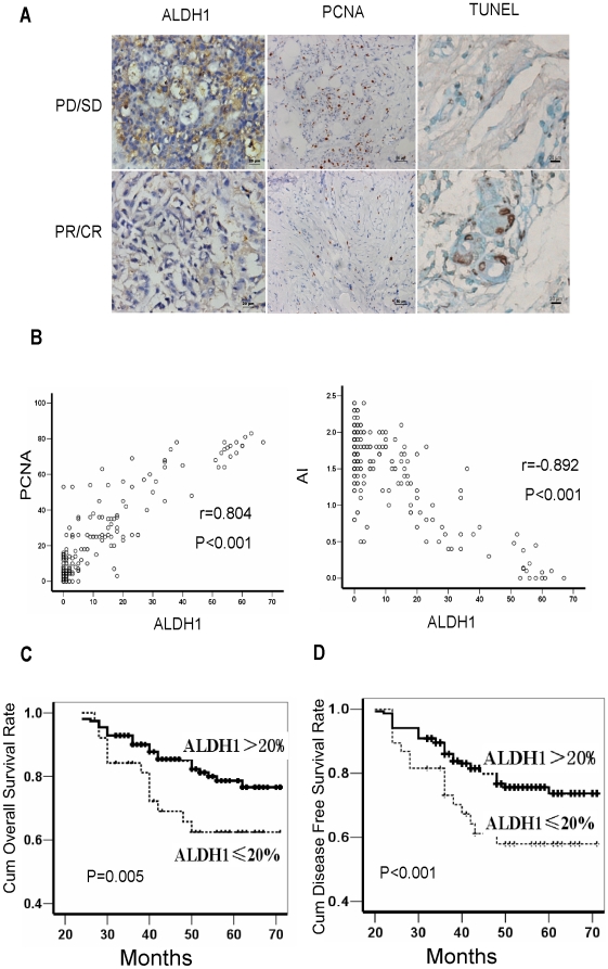
Figure 1. Aldehyde dehydrogenase (ALDH1) expression correlates with clinical outcome of breast cancer patients.
(A) Immunohistochemical staining shows tumors with poor clinical response (progressive or stable disease, PD/SD) to neo-adjuvant chemotherapy express high ALDH1 (>20% positive cancer cells) in pre-chemotherapy samples, and tumors with partal response (PR) express low ALDH1 (≤20% positive cancer cells). High proliferating cell nuclear antigen (PCNA)(>25% positive cancer cells) and poor apoptosis are observed in tumors with PD/SD after neo-adjuvant chemotherapy. Representative images of ALDH1 (×200), PCNA (×200) and Terminal deoxynucleotidyl transferase (TdT)-mediated dUTP labeling (TUNEL,×400). (B) Surgically removed tumor samples obtained after neoadjuvant chemotherapy from patients with low ALDH1 have higher percentage of TUNEL-staining cells (r = −0.892, p<0.001), but lower percentage of PCNA-staining cells (r = 0.804, p<0.001). (C–D)Kaplan-Meier curves with log rank tests show statistical difference in overall survival (Log Rank = 7.987, p = 0.005) (C) and disease-free survival (Log Rank = 19.347, P<0.001) (D) between patients with high ALDH1 expression and low ALDH1 expression.

