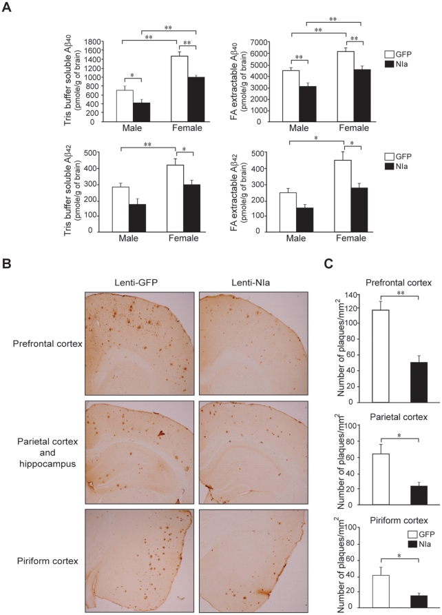
Figure 7. NIa-mediated reduction in Aβ levels and Aβ plaques in APPsw/PS1dE9 mouse brains.
(A) Brains of APPsw/PS1dE9 were bilaterally infused with Lenti-GFP and Lenti-NIa, and the amounts of Aβ1−40 and Aβ1−42 were measured by ELISA. The amounts of soluble and insoluble Aβ1−40 are shown (upper lane). The amounts of soluble and insoluble Aβ1−42 are shown (lower lane). (B) Sections of prefrontal cortex, parietal cortex, hippocampus, and piriform cortex of APPsw/PS1dE9 male mouse infused with Lenti-GFP and Lenti-NIa were stained with anti-Aβ antibody (Bam-10). (C) The number of plaques in the prefrontal cortex, parietal cortex, and piriform cortex of APPsw/PS1dE9 male mouse infused with Lenti-GFP and Lenti-NIa was counted. For Lenti-GFP infusions, n = 5 for male and n = 5 for female. For Lenti-NIa infusions, n = 6 for male and n = 3 for female. Error bars represent SD. *p<0.05 and **p<0.01.

