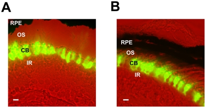
Figure 1. XCLΔQ-GFP expression in the specific cell-types in the transgenic photoreceptors.
A. An image of a section from the XOP-XCLΔQ-GFP tadpole retina. XCLΔQ-GFP is observed only in the cell bodies and inner segments of the rod photoreceptor cells. B. In a CAR-XCLΔQ-GFP transgenic retina, GFP accumulates only in the cone photoreceptor cells. CB: photoreceptor cell body; OS: photoreceptor outer segment; IR: inner retina; RPE: retinal pigment epithelium. The scale bar indicates 10 µm.

