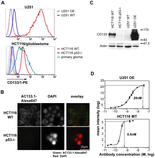
Figure 1. Characterization of cell lines and the AC133.1 antibody.
(A) CD133 expression of HCT116, U251, and primary glioblastoma stem-like cells as determined by flow cytometry. Upper panel: U251 wild-type cells and CD133-overexpressing U251 cell lines; lower panel: HCT116 wild-type, HCT116 p53−/−, and primary glioblastoma stem-like cells. Cells were incubated with the CD133/1-PE antibody and analyzed by flow cytometry. The dotted lines show the unstained controls. (B) CD133 expression of HCT116 wild-type and HCT116 p53−/− cell lines as determined by immunofluorescence microscopy. After incubation with the AC133-Alexa647 antibody, cells were counterstained with DAPI. (C) Levels of total cellular CD133 protein as determined by Western blotting. Actin served as a loading control. (D) Determination of the saturation concentration of AC133.1 antibody for HCT116 wild-type or CD133-overexpressing U251 cells via flow cytometry. The cells were stained using serial dilutions of the purified antibody and analyzed by flow cytometry using an anti-mouse PE-conjugated F(ab')2 fragment. The saturation concentrations were determined using prism software. WT, wild-type; OE, overexpressing.

