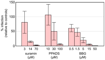
Figure 1. Inhibition of HDV infection by suramin, PPADS and BBG.
PHH were exposed to HDV in HGM for 3 h. From −1 to +3 h the inhibitors were also present, at the concentrations indicated. At +3 h both inhibitors and virus were removed and replaced by media containing preS1 peptide (50 nM) for the next 16 h, after which the cells were incubated in HGM out to 6 days, at which time total RNA was extracted and assayed by qPCR for HDV antigenomic RNA. As described in Methods, the mean values obtained are expressed relative to untreated control cultures. Error bars represent calculated standard error of the mean.

