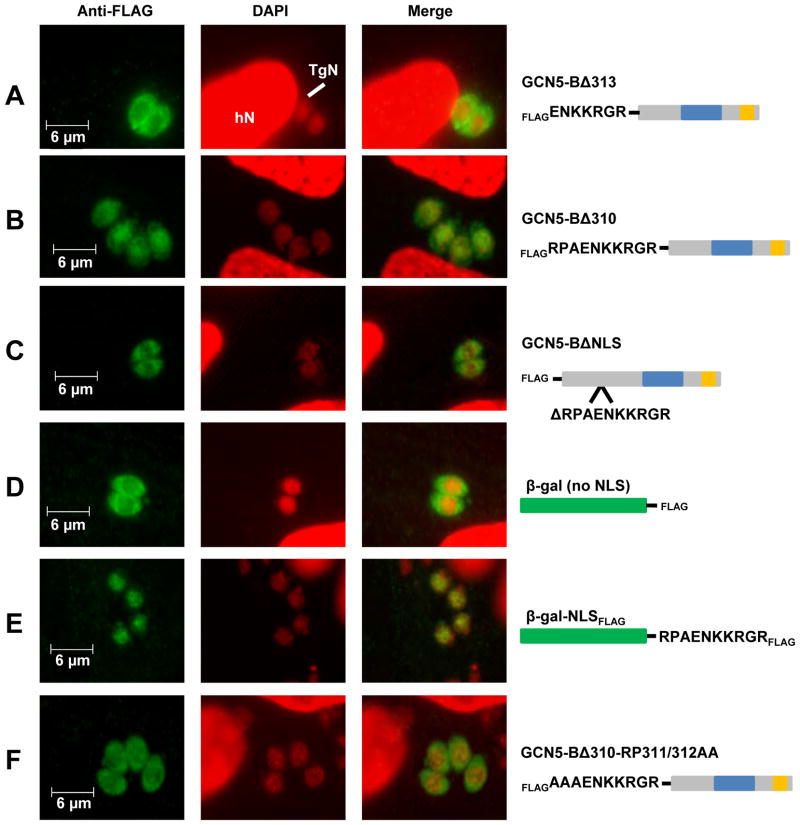
Figure 2. Mapping of the NLS for TgGCN5-B.
IFAs using antibody to FLAG tag were used to detect various forms of TgGCN5-B or β-gal fusion proteins. Diagram of each protein is shown to the right with FLAG epitope tag and proteins domains indicated. The blue box of each TgGCN5-B protein diagram represents the HAT catalytic domain and the orange box depicts the bromodomain. β-gal protein cartoon is represented in green. A. Localization of TgGCN5-B lacking the first 313 (Δ313) or B. 310 (Δ310) amino acid residues. C. Localization of TgGCN5-B after an internal deletion of the ten residue NLS (ΔNLS). D. Localization of β-galFLAG. E. Localization of β-gal-NLSFLAG. F. Localization of GCN5-BΔ310 containing alanine substitutions for R311 and P312. hN, host cell nucleus; TgN, Toxoplasma nucleus; green = anti-FLAG; red = DAPI.

