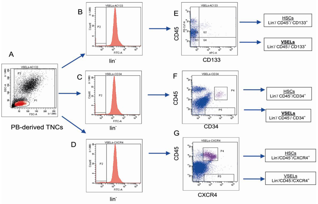
Fig. 1. Gating strategy for isolating VSELs by fluorescence- activated cell sorting (FACS).
Human VSELs were isolated from umbilical cord blood (CB)-derived total nucleated cells (TNCs) stained for: i) CD45 panleukocytic antigen, ii) hematopoietic lineages markers (Lineage/ Lin) and iii) CD133 stem cell antigen. Based on the size predefined beads (standard diameters of 1, 2, 4, 6, 10, and 15 µm) the gate R1 was set up to include all objects larger than 2µm (Panel A). TNCs were further visualized on the dot plot including region R1 and showing forward scatter (FSC) vs. side scatter (SSC) signals of cells that are related to the size and granularity/complexity of the cell, respectively (Panel B). Cells from region R1 were analyzed for hematopoietic lineages markers expression and only Lin− events were included into region R2. Population from region R2 was subsequently visualized based on CD45 and CD133 antigens (Panel D). Lin−/CD45+/CD133+ cells (VSELs) were sorted as events enclosed in logical gate including regions R1, R2 and R4, while Lin−/CD45−/CD133+ hematopoietic stem/progenitor cells (HSPCs) from gate including regions R1, R2 and R3. Percentages show the average content of each cellular subpopulation (± SEM) in total nucleated cells.

