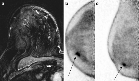
Fig. 2.
A 62-year-old female patient presented with a 1.8-cm ill-defined solid mass at 4 o’clock in the right breast by mammography that was diagnosed at pathology to be invasive mammary carcinoma with lobular features (grade I). MRI failed to detect any suspicious lesions in the right breast (a). b, c On PEM, a focal area of increased FDG activity corresponding to the biopsy-proven malignancy in the right breast and the inner lower quadrant approximately 1 cm from the nipple and measuring approximately 1.3 × 0.8 × 0.5 cm was clearly observed (arrows)

