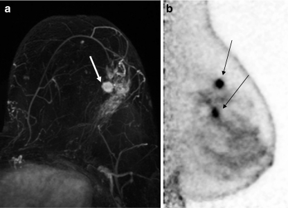
Fig. 3.
A 61-year-old subject presented with an area of clustered pleomorphic microcalcifications in the upper outer quadrant of the left breast which proved to be DCIS following stereotactic biopsy. An ultrasound confirmed the presence of the biopsy clip and identified an additional solitary 7-mm nodule in the same quadrant. Core sampling found this nodule to be IDC. MRI identified a 1.2-cm irregular enhancing mass (depicted by arrow) with a possible satellite lesion (a). PEM confirmed a 1.1 × 1.0 × 2.2-cm mass with a second 0.7 × 0.7 × 2.5-cm inferior mass with final pathology confirming IDC and DCIS, respectively (b), as depicted by arrows

