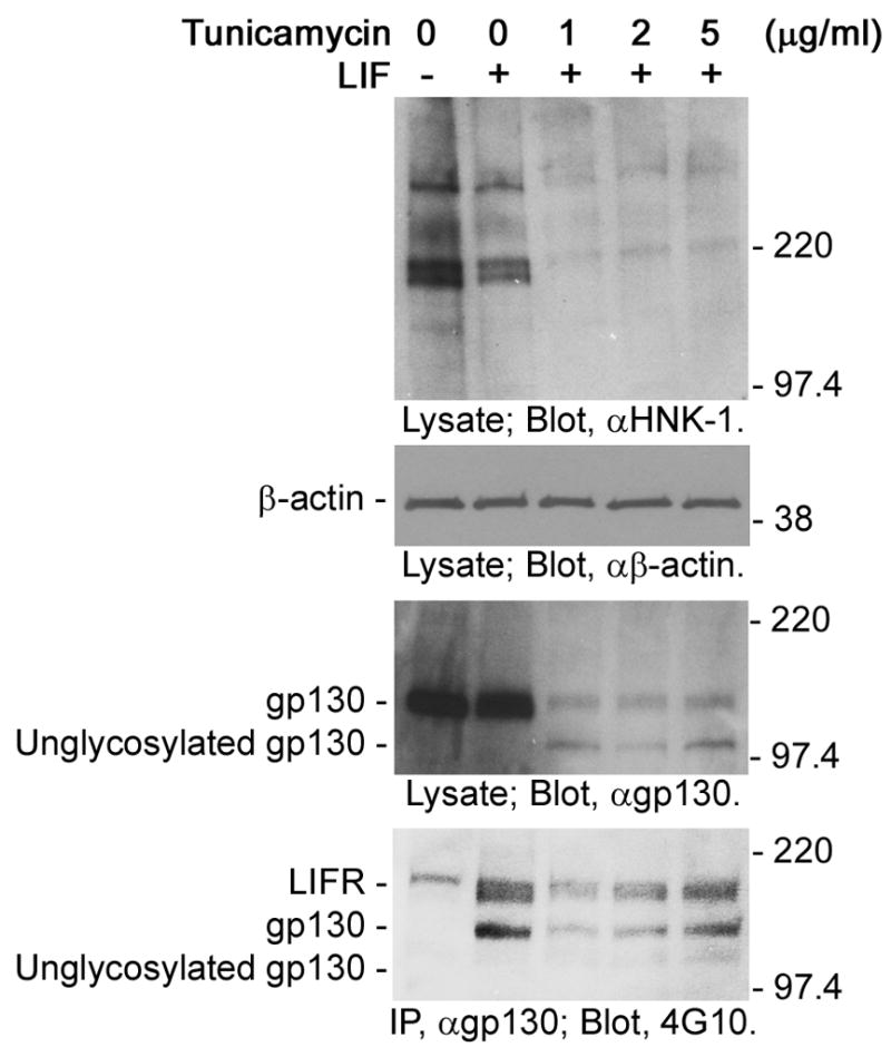
Fig. 1.

Unglycosylated gp130 in NECs treated with tunicamycin. NECs were treated with tunicamycin for 8 h, stimulated with LIF (0 or 40 ng/ml) for 10 min and then lyzed. Lysates and immunoprecipitated gp130 were subjected to Western-blot analysis. NEC lysates were analyzed with anti-HNK-1 antibody, anti-β-actin antibody and anti-gp130 antibody. Anti-HNK-1 antibody was used to detect glycoproteins as a control. Anti-β-actin antibody was used to confirm that the same amount of proteins was applied to each lane. Immunoprecipitated gp130 was analyzed with anti-phospho-tyrosine antibody (4G10) to evaluate the activation. The molecular masses are indicated on the right of each panel.
