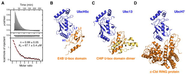
Figure 4. Interaction of E4B U-box with UbcH5c and Ubc4 E2 Conjugating Enzymes.
(A) Isothermal titration calorimetry of UbcH5c with E4B U-box. Shown are the integrated heat measurements from injecting 3 mM E4B U-box into the calorimeter cell containing UbcH5c at an initial concentration of 100 μM (top panel) or buffer solution (middle panel). A standard one-site model was used for curve fitting (bottom panel) in the determination of KD and stoichiometry (n), the values of which are shown.
(B) Crystal structure of human E4B U-box in complex with UbcH5c.
(C) Crystal structure of mouse CHIP U-box in complex with Ubc13 (PDB entry 2C2V).
(D) Crystal structure of the human c-Cbl RING E3 ligase in complex with UbcH7 (PDB entry 1FBV).
See also Figure S2.

