
Figure 8.
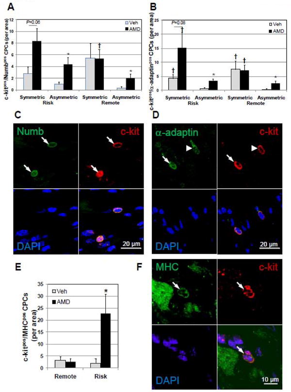
c-kitpos CPCs divide predominantly by symmetric mitotic division in AMD3100-treated hearts after ligation. The balance between self-renewal and commitment during cell division of the c-kitpos CPC pool was assessed by determining whether cell division was symmetric, indicative of self-renewal, or non-symmetric, indicative of the generation of a committed cell based on the polarized expression of Numb and α-adaptin. Myocardial commitment was assessed in c-kitpos CPCs by counterstaining for sarcomeric MHC and CPCs expressing both counted in risk and remote areas. Symmetric localization of Numb (A) and α-adaptin (B) in c-kitpos CPCs in AMD3100-treated hearts after ligation is increased in the risk area. (C and D) Representative confocal images of c-kitpos CPCs displaying symmetric (arrows) and asymmetric (arrow head) localization of Numb and α-adaptin. (E) Myocardial committed CPCs, c-kitpos/MHCpos are significantly elevated in AMD3100-treated infarcted hearts. (F) Representative confocal images of c-kitpos/MHCpos CPC (arrow). Values are mean ± SEM (n =6). *P<0.05 vs Vehicle; †P<0.05 vs Asymmetric.